Types of Ear Deformities
Newborn ear deformities occur in roughly one-third of infants, sometimes resolving on their own but often requiring medical treatment to restore a natural-looking appearance to the ears. As children with ear deformities grow older, the misshapen ear anatomy could cause problems with hearing as much as aesthetic concerns. Fortunately, an array of the most common ear-related deformities can be corrected with non-surgical treatment to reshape the ears, repair any auditory issues, and achieve a conventional ear appearance. Our experienced craniofacial plastic surgeon, Dr. Eric Payne, offers the most advanced non-surgical and surgical ear reshaping procedures available to correct these concerns.
Follow the links below to learn more about the various types of newborn ear deformities. If you have additional questions, or if you would like to schedule a consultation, please contact our office today.
What are the Types of Ear Deformities?
There are several different types of ear deformities commonly found in children. An ear deformity is distinct from an ear malformation, which is a condition that involves missing skin and cartilage. A newborn with an ear deformity, on the other hand, is not missing any ear skin or cartilage, but experiences misshapen ear(s). Typical ear malformations include microtia or anotia, meaning the child is born with one ear or the ear(s) are partially/completely missing. Cryptotia is also considered an ear malformation since the condition often involves missing skin.
Many different kinds of ear deformities can be repaired with non-surgical infant ear molding. Treatable conditions using this technique include:

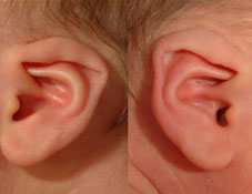
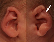
Stahl’s Ear Deformity
Sometimes called Spock ear, Vulcan ear, elf ear, or Satyr ear, this condition occurs when the upper-third of the ear has a pointed or prominent shape. In some cases, this is due to an extra fold in the helix, a flattened section of the helical rim, or both. With Stahl’s ear deformity, the upper part of the ear may stick out from the scalp.
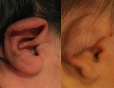
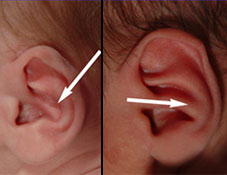
Lidding Ear Deformity
Similar to lop ear deformity, this condition causes the upper helical rim of the ear to fold over. Generally, lidding ear deformity will involve a tighter crease than a lop ear deformity and there may even be a rolled appearance in severe cases.
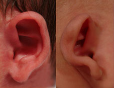
Lop Ear Deformity
Lop ear is defined as a folding of the upper-third or half of the ear. This newborn ear deformity is very similar to lidding in that the upper helical rim is folded over.
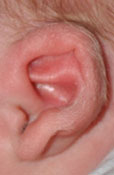
Prominent Ear Deformity
Projection of the mid-rim from the skull greater than 7 mm is defined as a prominent ear deformity. This is considered the most common defect, accounting for 45% of all ear deformities. It is frequently overlooked in infants and very difficult to treat, especially with surgery. A variation of prominent ears is called “Cup Ear,” which is its most severe form. Some have suggested a genetic link, as parents with prominent ears frequently have children with the same deformity; however, the true cause of prominent ears is abnormal intrinsic ear muscles, which create bends or folds in the ears. When the muscle is missing or in an irregular position, a prominent ear deformity can result.
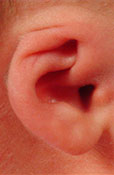
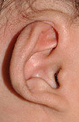
Cup Ear Deformity
In this advanced prominent ear deformity, the helix and lower lobe (lobule) of the ear are pushed forward and bend in toward each other and toward the head, while the middle portion of the ear remains back. This creates a cup-like shape. It’s considered a prominent ear deformity, as it causes the ear to project significantly outward from the head.
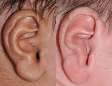
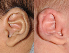
Constricted Ear Deformity
Constricted ear deformity refers to a number of defects wherein the helical rim is folded, wrinkled, or very tight. The ear often experiences reduced height due to the folded or flattened appearance. The constricted ear deformity is best described as tight skin around the cartilage, restricting a normal shape.
Conchal Crus Deformity
The conchal bowl is made up of two parts: the cymba superiorly and the cavum inferiorly. The conchal crus ear deformity is an abnormal outward fold dividing the concha cymba in half. This deformity can be corrected using the EarWell™ device with the conchal conformer or a custom ear mold. Conchal crus appears similarly to a Boxer’s ear or cauliflower ear.
Inverted Conchal Bowl Deformity
The conchal bowl is the hollow or depression located near the ear canal. It is made up of two indentations called the cymba and cavum. In the inverted conchal bowl deformity, the concha is projected outward, causing a narrowing of the external ear canal and possible changes in the anti-tragus of the lower ear. This can later cause problems using headphones or earbuds, even leading to issues with ear wax buildup in the ear canal. The inverted conchal bowl deformity can also reduce hearing function on the affected side. It was first described and illustrated as a Mozart ear deformity.
Cryptotia
Also called buried ear or hidden ear, cryptotia is a defect in which the upper ear is partially concealed by the side of the head. In many cases, the scaphoid fossa and the antihelical crura also will be smaller and underdeveloped, giving the ear a pinched appearance. These areas are surrounded by the helix of the upper ear. Typically, skin flaps or grafts are required to correct this deformity with surgery. The biggest problem is often the inability to wear eyeglasses or sunglasses since the ear is missing a normal skin fold and projection. Ear molding has been successful in eliminating the need for future surgery with skin grafts.
Helical Rim Ear Deformity
This defect occurs when there is a lack or deficiency of skin/cartilage in between the scapha and the rim, causing a clipped, depressed, or indented appearance on the outer edge of the upper ear.
Mixed Ear Deformity
Mixed deformity of the ear involves multiple conditions combined, such as Stahl’s, lidding, lop, constricted, or other defects. The EarWell™ device can effectively correct a number of common and complex infant ear deformities—including mixed ear deformities—if utilized soon after birth. Every patient’s situation is unique, and Dr. Payne will discuss the optimal treatment plan during the initial consultation.
What Causes Ear Deformities?
Ear deformities are usually present at birth (congenital) or formed through trauma or injury. It is theorized that an infant’s position in the womb or other environmental factors prior to birth may factor into why an ear deformity occurs, but every newborn differs and it is possible for an ear anomaly to be completely random. Another key factor that may cause an ear anomaly is missing or abnormal positioning of the ear muscles to the cartilage, which can change the shape and folding of the ear. Ear deformities can also be inherited or caused by a genetic mutation.
While it is not always possible to diagnose an ear deformity before birth, potential conditions can typically be detected through a physical exam. This may involve a hearing test and/or a digital imaging scan. The outlook for non-surgical correction of an ear deformity is usually very positive if the condition is diagnosed within a few weeks of birth.
How are Ear Deformities Repaired?
Traditionally, parents were typically advised to wait until their child reached five to seven years of age to surgically correct their ear deformity. This often forced children to contend with being teased by their peers and classmates throughout childhood. Now, many types of ear deformities can be repaired non-surgically within a few weeks of birth. The EarWell® Infant Ear Correction System is a device designed to gently mold the ears into the proper shape without the need for incisions, surgery, or downtime. EarWell® boasts a 90 percent success rate when the device is used within the first three weeks of life and results are generally more effective the earlier the system is utilized.
EarWell® uses small retractors and conformers to remodel the cartilage of the ear into a conventional ear shape. These ear molding pieces are held in place with an anterior shell placed over the ear, allowing the ear to conform to the desired curvature. The EarWell® device is left in place for two weeks at a time and changed for a treatment course of six to eight weeks on average depending on how young your child is and the type of deformity being treated. Dr. Payne has helped countless infants achieve a normal ear appearance using the EarWell® system. Having received training from Dr. Steve Byrd himself, the inventor of EarWell®, Dr. Payne has the experience to evaluate your child’s concerns and recommend the most effective treatment option according to their needs. You can also check with your pediatrician to learn whether your baby is a candidate for ear molding.
Contact Us
For more information about the various types of ear deformities, please contact our office today. Our skilled medical team can answer your questions or help you to schedule a consultation.